
How Mercury is Transferred to the Human Body
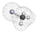
Mercury is most harmful to humans in the form of methylmercury. Methylmercury is a an organometallic cation in the form of H3C-Hg+. The positive charge on methylmercury ion warrants the molecule to interact with other negatively charged ions. Methylmercury has a high affinity for binding to proteins that contain the amino acid cysteine.
After ingestion of food containing methylmercury, such as fish, the cation is absorbed into the blood stream through the gastrointestinal tract and transported throughout the body. While traveling in the blood, methylmercury is free to interact with other proteins and is often found bound to cysteine containing proteins. The combination of methylmercury and cysteine creates a compound that is similar in structure to methionine. Amino acid transport proteins are embedded in the blood-brain barrier and placenta. The transport protein that is responsible for moving methionine across the blood-brain barrier and the placenta are fooled by the methylmercury-cysteine complex and shuttle it into the brain and placenta where it can then cause serious health complications.
After ingestion of food containing methylmercury, such as fish, the cation is absorbed into the blood stream through the gastrointestinal tract and transported throughout the body. While traveling in the blood, methylmercury is free to interact with other proteins and is often found bound to cysteine containing proteins. The combination of methylmercury and cysteine creates a compound that is similar in structure to methionine. Amino acid transport proteins are embedded in the blood-brain barrier and placenta. The transport protein that is responsible for moving methionine across the blood-brain barrier and the placenta are fooled by the methylmercury-cysteine complex and shuttle it into the brain and placenta where it can then cause serious health complications.
Mercury's Effect: Unborn Babies, Infants, and Small Children

It has been medically determined that children are most vulnerable to mercury poisoning--affecting both small children and developing fetuses in the womb. The developing fetus is exposed to mercury because it is easily transported across the placenta along with other essential nutrients. The first two months of gestation are the most critical time for proper development of the brain and nervous system and mercury exposure should be closely monitored during this time.
Maternal exposure to mercury is considered dangerous both before and during pregnancy since mercury stays in the mother’s body for an extended time before it is slowly excreted. Mercury is a potent neurotoxin and once the fetus has been exposed to it, it can damage, destroy, or impair nervous tissue function. As a result, this directly affects development of the baby’s brain and nervous system. Some of the symptoms of mercury exposure may not show right away in the newborn infant, but later present as learning difficulties once a child is enrolled in school. For an example, young children can have compromised vision and difficulties hearing, stunted motor skills, impeded language development, decreased IQ, problems with memory, and attention deficit.
Maternal exposure to mercury is considered dangerous both before and during pregnancy since mercury stays in the mother’s body for an extended time before it is slowly excreted. Mercury is a potent neurotoxin and once the fetus has been exposed to it, it can damage, destroy, or impair nervous tissue function. As a result, this directly affects development of the baby’s brain and nervous system. Some of the symptoms of mercury exposure may not show right away in the newborn infant, but later present as learning difficulties once a child is enrolled in school. For an example, young children can have compromised vision and difficulties hearing, stunted motor skills, impeded language development, decreased IQ, problems with memory, and attention deficit.
Brain Development in Babies and Children

The effects of mercury exposure on human brain development begins during the formation and organization of brain cells in the developing embryo. The fully formed human brain--composed of approximately 100 billion nerve cells--is developed three to four weeks after conception. Throughout pregnancy, the production of nerve cells is so fast nearly half of the human genetic code is dedicated to producing the brain itself. Genetics determine the majority of human brain development but environmental factors also play a role. Included in these factors are the health and nutritional state of the mother and her contact with toxic substances such as alcohol, tobacco, drugs, and mercury--all of which affect healthy brain development.
Numerous studies confirm that mercury in particular changes the function and structure of the central nervous system by decreasing the electrical activity across the nerve cell connectors called synapses. By decreasing this essential electrical activity of the brain, it prevents the release of important neurotransmitters such as dopamine. Dopamine is very important for brain functions such as attention, thinking and motor skills. Research has proven that prenatal exposure to mercury resulted in a loss of motor skills and control during the most important development stage: when dopamine nerve cells fully grow into functioning brain cells. Mercury also affects the connectivity of these nerve cells resulting in a reduction of functioning brain cells, brain size and overall brain weight.
Numerous studies confirm that mercury in particular changes the function and structure of the central nervous system by decreasing the electrical activity across the nerve cell connectors called synapses. By decreasing this essential electrical activity of the brain, it prevents the release of important neurotransmitters such as dopamine. Dopamine is very important for brain functions such as attention, thinking and motor skills. Research has proven that prenatal exposure to mercury resulted in a loss of motor skills and control during the most important development stage: when dopamine nerve cells fully grow into functioning brain cells. Mercury also affects the connectivity of these nerve cells resulting in a reduction of functioning brain cells, brain size and overall brain weight.
How to Protect Your Baby and Children from Mercury Exposure
The risks of mercury exposure are highest in the unborn fetus where the developing baby receives 100 percent of its nourishment from its mother. However, infants and small children are also at risk through their diet. Infants are still at high risk to mercury exposure by consuming breast milk, and since small children tend to eat more food than adults in proportion to their body size, a large serving of fish tainted with mercury will affect the health of a small child much more than it would an adult. Mothers can be exposed in a variety of ways: inhalation, consumption of food or water, or by receiving a vaccine. However, the primary source of mercury exposure in the United States is from eating fish and shellfish contaminated with mercury in their flesh. This is why it is very important that both pregnant women and mothers of infants and small children take action by limiting their consumption of fish known to contain high levels of mercury.
References:
↜ PRevious page | Next page ↝
Created by Portland State Multimedia Capstone Winter 2015
